
Jarvis
Dr. Balaji Bashyam's AI Assistant
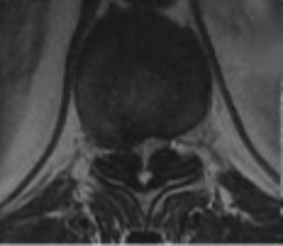
Ossified yellow ligament — also called ossification of the ligamentum flavum (OLF) — is a condition where the yellow ligament at the back of the spinal canal slowly turns to bone. As it thickens, it can press on the spinal cord, most often in the chest (thoracic) region, leading to unsteady walking and leg weakness. This guide explains the warning signs, how it is diagnosed on CT and MRI, and the surgical options that relieve the pressure.
In short: Ossified yellow ligament is a common cause of thoracic spinal cord compression. There is no tablet or injection that reverses the bone formation. When it causes myelopathy (spinal cord dysfunction), the effective treatment is surgical decompression — removing the ossified ligament to free the spinal cord. This can be done through open, microscopic, or (in selected cases) endoscopic techniques.
The ligamentum flavum (Latin for “yellow ligament”) is an elastic band that lines the back wall of your spinal canal and helps hold the vertebrae together. In some people it gradually calcifies and then ossifies — genuine bone forms within it. This is called ossified yellow ligament (OYL) or ossification of the ligamentum flavum (OLF).
Because the ossified ligament sits directly behind the spinal cord, it narrows the canal (spinal stenosis) and squeezes the cord from behind. It occurs most commonly in the lower thoracic spine, but can appear anywhere from the upper thoracic to the thoraco-lumbar junction. It develops slowly, so symptoms can creep in over months to years and are often mistaken for age, arthritis, or a “slipped disc.”
Because the pressure is usually in the chest region of the spine, symptoms typically appear in the legs and trunk rather than the arms. Watch for:
Symptoms that are progressing — especially worsening balance or new bladder trouble — should be evaluated promptly, because earlier treatment gives better recovery.
Two scans are used together, because each shows something different:
MRI is the key test for the spinal cord. It shows how severely the cord is being compressed and whether it has been injured — a bright (high-signal) area within the cord on T2 images suggests cord strain (myelomalacia) and helps decide how urgently to operate. Note that the ossified ligament itself is bone, so it appears dark on MRI and can be easy to miss; MRI shows the effect (the pinched cord) rather than the ossification directly.
A CT scan shows the bone in fine detail — the exact size, shape and level of the ossified ligament. This is essential for planning the safest surgical approach, and for spotting whether ossification is present at more than one level or on both the front and back of the canal.
Why both scans matter: MRI answers “how badly is the cord affected?” and CT answers “exactly where and how much bone is there?” Together they let your surgeon tailor the operation precisely to your anatomy.


Once the ossified ligament is causing spinal cord symptoms, medication and physiotherapy can support recovery but cannot remove the bone or reverse the compression. The definitive treatment is surgical decompression. The goal is the same in every case — take the pressure off the spinal cord — but the technique is matched to how extensive the ossification is and where it sits.
This is the established, time-tested approach, especially when the ossification spans several levels or is large. The surgeon removes the bony back wall of the canal (laminectomy) along with the ossified ligament, giving the cord room to recover. In selected patients, screws and rods are added to keep the spine stable and correctly aligned after decompression.
Here the same decompression is performed under a high-powered surgical microscope. The magnification and bright, focused light allow the ossified ligament to be peeled away from the delicate cord with great precision, which is valuable because the ossified ligament is often stuck to the covering of the cord.
For focal, single-level or limited ossification in suitable patients, a minimally invasive endoscopic technique can be used. Working through a small port with a high-definition camera, the surgeon removes the ossified ligament through a much smaller incision. Potential advantages include less muscle disruption, less blood loss, and quicker early mobilisation. Endoscopic surgery is not appropriate for every case — very extensive or circumferential ossification is usually safer through an open or microscopic route — so careful case selection is essential.
Occasionally the yellow ligament ossifies at the same time as the posterior longitudinal ligament (OPLL) at the front of the canal, so the cord is squeezed from both sides at once — “circumferential” compression. This is rare and technically demanding. A recent case report described a 45-year-old woman with rapidly worsening walking due to combined ossified yellow ligament and OPLL at the upper thoracic spine (T3–T4). Rather than the higher-risk approach of opening the chest from the front, the surgeons used a carefully planned posterior-only strategy — laminectomy, removal of the ossified yellow ligament, a costotransversectomy-assisted release of the front ossification, and stabilisation — and achieved good decompression. The patient improved meaningfully over three months (Nurick grade 5 to grade 3).1 The lesson for patients: even complex, dual-ligament compression can often be managed through a single well-designed posterior operation, avoiding the risks of anterior chest surgery.
The main aim of surgery is to stop the condition getting worse and give the spinal cord the best chance to recover. Patients who have decompression at the right time can expect:
Recovery is usually gradual over weeks to months and is supported by physiotherapy. The single biggest factor in how much you recover is how early the cord is decompressed — long-standing, severe compression recovers less completely, which is why prompt assessment matters.
Unsteady walking, leg weakness or numbness that is slowly getting worse should never be ignored. Dr. Balaji Bashyam, Consultant Endoscopic & Minimally Invasive Spine Surgeon, evaluates and treats thoracic cord compression and ossified yellow ligament, offering open, microscopic and endoscopic decompression matched to your condition. Consultations are available in Chennai (Choolaimedu & Aminjikarai) and at visiting clinics in Nellore, Tirupati and Thiruvannamalai.
📞 Book an appointment: +91 90031 85183
Is ossified yellow ligament the same as a slipped disc?
No. A slipped (herniated) disc is soft disc material pressing on nerves, usually in the neck or lower back. Ossified yellow ligament is actual bone forming in a ligament at the back of the spinal canal, most often in the chest (thoracic) region, and it presses on the spinal cord itself. The symptoms and treatment are different.
Can ossified yellow ligament be treated without surgery?
There is no medicine or injection that dissolves the ossified bone or reverses the compression. If there are no cord symptoms, it may simply be monitored. But once it causes myelopathy — unsteady walking, leg weakness, numbness or bladder changes — surgical decompression is the effective treatment.
Is surgery for ossified yellow ligament safe?
Decompression is a well-established operation performed routinely by spine surgeons. As with any spinal cord surgery there are risks, which your surgeon will explain, but modern techniques — including microscopic and endoscopic methods — allow the ossified ligament to be removed precisely. Careful planning with MRI and CT, and operating before the cord is severely damaged, keep outcomes favourable.
Can ossified yellow ligament be removed by endoscopic (keyhole) surgery?
In selected patients with focal, single-level or limited ossification, yes — endoscopic decompression can remove it through a small incision with less muscle damage and faster early recovery. Very extensive or front-and-back (circumferential) compression is usually safer through open or microscopic surgery. Your surgeon decides based on your scans.
Will I be able to walk normally again after surgery?
Many patients regain steadier walking and better strength, especially when surgery is done before the cord is badly damaged. Recovery is gradual over weeks to months and is helped by physiotherapy. How much you recover depends largely on how severe and how long-standing the compression was before surgery.
How quickly should ossified yellow ligament be treated?
If symptoms are progressing — worsening balance, increasing leg weakness, or any new bladder or bowel difficulty — it should be assessed promptly. Early decompression protects the spinal cord and gives the best chance of a good recovery.
This article is for general education and does not replace an in-person medical consultation. If you have progressive weakness, numbness or bladder symptoms, please seek assessment by a qualified spine surgeon.